Welcome to Cherry Times




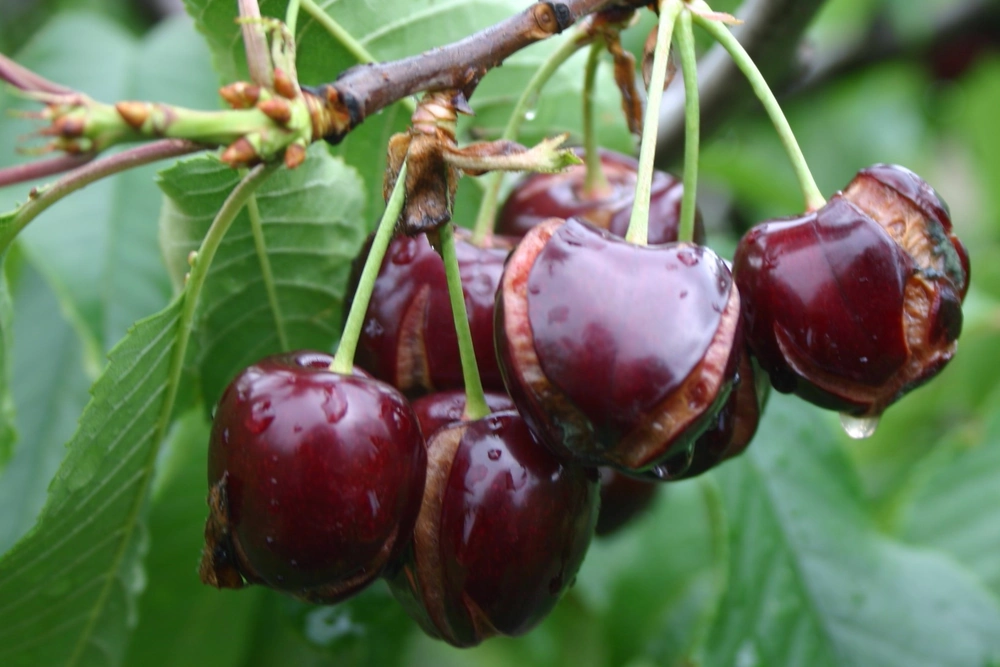 Image source: Stefano Lugli
Rain exposes a vulnerability that develops in cherries as they ripen. To understand cracking, we need to follow the fruit’s growth, the changes in its skin, and what happens when water reaches a weakened area. Current knowledge places the start of the damage at a local level and links its spread to a loss of tissue cohesion. This perspective helps explain why apparently similar fruit can respond differently to the same exposure to water.
As the cherry grows, its surface area must increase along with the volume of its flesh. Its growth follows a double-sigmoid curve: after an initial period of active growth, pit hardening coincides with a slowdown in the increase in fruit size; later, cell expansion in the mesocarp drives the final growth phase. During this third phase, the surface begins expanding rapidly again, but the formation of the outer covering can no longer keep pace (Knoche et al., 2004; Alkio et al., 2012).
This covering is the cuticle, a matrix of cutin and waxes deposited on the epidermis. Its formation is concentrated in the early stages and declines before the final expansion phase. As a result, a quantity of material that increases only slightly must cover an increasingly large surface: the amount of cuticle per unit of surface area decreases, and strain builds up.
Deposition
The amount of cuticular material covering the fruit surface.
Strain
How much the surface has expanded relative to its relaxed state.
These two phenomena describe distinct aspects of the cuticle (Knoche et al., 2004).
The decline in deposition also has a biological dimension. The expression of candidate genes involved in the synthesis and transport of cuticular components follows the development of the cuticle as the fruit grows. By the final expansion phase, the fruit therefore has a much less active cuticle-formation program. This correspondence links gene regulation to the construction of the barrier, but coordinated expression alone does not show which gene determines resistance to cracking (Alkio et al., 2012).
The result is a surface increasingly prone to developing microcracks. These discontinuities may remain confined to the cuticle and be invisible to the naked eye. A fruit that appears commercially intact may therefore have already lost some of its protection against water, without yet showing a crack in its tissues (Knoche and Peschel, 2006).
A microcrack does not mean that the entire skin has ruptured, because the skin is a composite structure. Beneath the cuticle are the epidermis and hypodermal layers, whose cell walls bear much of the mechanical load. Biaxial tensile tests, which stretch skin segments in two directions, indicate that these tissues provide the main structural support; the cuticle’s contribution is relatively small (Brüggenwirth et al., 2014).
This structure links two challenges: maintaining the water barrier and holding together tissues under tension. The latter depends both on cell-wall strength and on adhesion between cells. Pectins in the middle lamella, located between the walls of adjacent cells, play a central role in this adhesion.
In cherry parenchyma, experimental pectin degradation makes cell separation easier, while microscopic markers help locate components of this matrix. The findings confirm its adhesive function, but they are not equivalent to measuring cracking resistance in different varieties (Schumann et al., 2019; Culemann et al., 2026).
Cohesion also depends on the hydration of the cell-wall matrix. In tests on cherry skin, greater hydration-driven wall swelling is associated with lower fracture resistance and more frequent cell separation. In addition, fractures in stretched segments can pass through cell walls, whereas cracking induced by immersion occurs predominantly between cells.
Therefore, stiffer skin, firmer flesh, and greater resistance to cracking are not interchangeable properties (Brüggenwirth and Knoche, 2017).
Hydration-driven cell-wall swelling and the increase in volume of a cell that absorbs water are distinct processes. In the first, the wall itself becomes thicker. In epidermal segments, loss of turgor allows this increase; restoring turgor through deplasmolysis can reverse it. Cell pressure therefore helps limit cell-wall swelling.
This local constraint is consistent with the low turgor of ripe fruit and does not imply a general increase in internal pressure as the cause of fracture (Schumann and Knoche, 2020).
When it rains, water encounters a barrier whose continuity and tension depend on the fruit’s previous development. Wetting can worsen microcracking in the strained cuticle. Exposure is also uneven: water may collect in the stem cavity or form a droplet at the stylar end. Thus, the wetted surface area and the duration of contact matter physiologically, as well as the total amount of rainfall (Knoche and Peschel, 2006; Winkler et al., 2020).
Water moves in response to differences in water potential, which describes its energy state. The accumulation of solutes makes osmotic potential more negative and promotes water entry when a transport pathway is available. A cherry, however, is not a uniform compartment: a more negative osmotic potential has been measured in the flesh than in the epidermal cells.
This variation helps explain how water is distributed within the fruit, but the osmotic difference does not directly measure either flow or pressure (Grimm and Knoche, 2015).
Pressure-probe measurements show low turgor in the mesocarp cells of ripe cherries. In addition, water-uptake tests have not detected the general pressure increase that would be needed to attribute cracking solely to internal overpressure caused by water entry. Skin tension remains relevant: cell pressure, osmotic status, and mechanical tension are different quantities (Knoche et al., 2014; Grimm et al., 2019).
The fruit also continues to exchange water with the tree. During development, estimated xylem inflow declines markedly, while the relative importance of phloem inflow increases. These estimates are based on changes in fruit diameter and selective interruption of transport; they do not justify claiming that xylem flow is always zero or that water management is irrelevant.
Instead, they require us to distinguish the fruit’s vascular water balance from localized water entry through a wetted surface (Brüggenwirth et al., 2016).
Wetting one area can cause cracking even when the fruit as a whole is losing water through transpiration. Similarly, immersion of harvested fruit and simulated rain on fruit still attached to the tree produce different responses. The total amount of water absorbed, by itself, does not show where water accumulates or which tissue loses integrity (Knoche and Peschel, 2006; Winkler et al., 2020).
To explain the damage, we need to look inside the fruit, at the cells beneath a discontinuity. Magnetic resonance imaging using labelled water located its entry in microcracked regions. Optical coherence tomography tracked, without cutting the fruit, the increase in volume of outer mesocarp cells near the point where a crack develops. Changes were much smaller in more distant areas.
These observations were made on harvested fruit immersed in water, but they provide direct evidence of where the process takes place (Grimm et al., 2019).
In these observations, cell expansion occurred before the crack was detected and intensified as it developed; the subsequent loss of volume is consistent with cells rupturing and emptying. The resolution of the experiment limits how precisely the sequence can be timed. Damage to a few cells may alter the immediate surrounding environment and promote further damage in skin that is already under tension (Grimm et al., 2019).
When cellular compartmentalization is lost, cell contents come into contact with nearby walls and membranes. Tests using juice and malic acid solutions show that this contact can promote cracking and loss of membrane integrity; much of the effect depends on acidity.
The findings support a local amplification mechanism, but they do not yet quantify the concentration or timing of acid release in a crack advancing during natural rainfall (Winkler et al., 2015).
Microscopy of the crack front shows cuticle discontinuities, cell death, hydration-driven wall swelling, and separation between cells beside intact tissue (Figure 1). The crack can advance along the middle lamella and extend into the flesh. The so-called “zipper” model brings these processes together: a localized lesion reduces cohesion and makes it easier for the damage to spread.
Experimental evidence supports the individual components of this model; the contribution of each may vary according to the tissue and exposure conditions (Schumann et al., 2019).
Editorial outline of the processes described in the text; it does not represent a universal timeline or quantify flows or pressures.
Figure 1. Two scales of wetting-induced cracking. A: areas where water may collect, without indicating a single point of origin. B: a microcrack limited to the cuticle and a crack reaching the underlying tissues represent distinct schematic stages. The blue cell highlights the outer mesocarp near the point where water enters.
Internal outlines and cell-wall boundaries are shown schematically; the reddish area beside the crack indicates local cell damage, without establishing exactly when it occurs. The blue arrows represent the localized entry of water and its inward progression observed in tests; they do not indicate a single microscopic pathway or quantify flow. The dashed arrow represents the hypothesized contribution of released cell contents to the weakening of the surrounding area.
The drawing does not establish a universal timeline between cell rupture and visible opening, and it does not depict pressure. Anatomy and thicknesses are simplified and not to scale. Original artwork based on Knoche and Peschel (2006), Brüggenwirth et al. (2014), Grimm et al. (2019), Schumann et al. (2019, 2020, 2022), Schumann and Knoche (2020), and Winkler et al. (2015, 2024).
Calcium helps explain the importance of cohesion. It can link pectin chains through their available carboxyl groups, so both the structure of the pectin and the surrounding environment matter. In cherry skin segments and extracted cell walls, adding calcium reduces hydration-driven wall swelling, while removing it with a chelating agent promotes swelling. pH also changes the response.
These tests confirm a relationship between the chemical environment and cell-wall stability, but on their own they do not demonstrate the entire chemical sequence that takes place in a crack under field conditions (Schumann et al., 2022).
In experiments involving direct contact with tissues and cracks, calcium also reduces the rate at which cracking spreads. However, increasing the total calcium content of the fruit through sprays has not always provided equivalent protection. This difference indicates that the location and availability of calcium at the damage site matter; the agronomic effectiveness of a treatment must be assessed separately (Winkler et al., 2024).
This sequence may begin and progress more or less easily depending on the genotype. Its heritable component is supported by genomic regions associated with cracking, identified both in populations monitored over several years under natural rainfall and in offspring assessed through immersion tests. The two approaches provide complementary information, but measure different exposure conditions.
A QTL identifies a genomic region associated with a trait; it does not automatically identify a causal gene or indicate tolerance that is independent of the environment (Quero-García et al., 2021; Crump et al., 2022).
The physiological question is how this inherited variation is expressed in the barrier and tissues. In one offspring population, certain cuticle characteristics, microcracking, and calcium status were linked to susceptibility. These findings connect differences between genotypes to specific processes, but they come from a single population and do not establish a universal indicator of resistance.
Susceptibility depends on a combination of properties, not on a single measurement of cuticle thickness or firmness (Knoche et al., 2025).
Some limited functional evidence is also beginning to emerge. Transient overexpression of PavCML42, a candidate gene linked to calcium signalling, increased cracking associated with the infiltration point in harvested cherries and changed the activity of cell-wall enzymes. This result helps link signalling to tissue disorganization, but the inoculation wound and experimental system limit how far it can be applied to intact fruit exposed to rain.
It does not yet provide a general explanation for differences between varieties (Yang et al., 2026).
As harvest approaches, fruit growth and cell-wall transformation occur together. Tissue expansion changes the proportions of structural materials. In a study tracking the ‘Regina’ variety, total cell-wall mass per fruit increased, but more slowly than fresh mass; the amount of cellulose per fruit remained almost constant, while its proportion of the cell wall declined.
As a result, a decrease in cell-wall material per unit of fresh mass does not necessarily mean that its absolute amount per fruit has fallen: it may also reflect differences in the rates of deposition and growth (Schumann et al., 2020).
At the same time, the composition of the matrix changes. In the same study, the water-soluble pectin fraction increased, and the distribution of other pectin fractions changed. The cell wall remains physically present, but the organization of components that contribute to cohesion and hydration changes. These fractions are defined by the solvents used to extract them: their quantities do not directly measure the strength of adhesion between cells.
To interpret cracking, these chemical changes must be linked to the tissue’s response, without assuming that an increase in soluble pectins directly increases cracking (Schumann et al., 2020; Culemann et al., 2026).
Comparing samples collected at different stages of development makes it possible to relate chemical changes to the physical properties of the cell wall. The increase in epidermal cell-wall thickness after turgor was removed was greater at later stages. However, the hydration-driven swelling capacity of extracted walls did not follow exactly the same pattern: it tended to stabilize during stage III.
An extracted cell wall loses some of the interactions and constraints it retains within the tissue. These findings show how its potential response changes during ripening, but they do not show that the walls have already swollen to this extent before rain falls (Schumann et al., 2020).
Hormonal regulation of development can also alter the fruit’s structural vulnerability. In a trial on the ‘Summit’ variety, applying abscisic acid (ABA) before the colour change increased cracking and was accompanied by anatomical changes and changes in the expression of cell-wall-related genes. The treatment provides evidence of a hormonal effect under those conditions, but it does not fully separate that effect from possible differences in ripening stage.
Changes in gene expression point to processes associated with the response; the causal role of each gene and the extent of ABA’s effect under other conditions remain to be determined (Liu et al., 2026).
To interpret what happens in the orchard, it helps to reconstruct the sequence: what condition the fruit was in before it got wet, where the water remained, and how the damage may have started and spread. Colour, size, and firmness provide information about some aspects of ripening, but on their own they do not reveal the microscopic continuity of the barrier or the tissue’s ability to maintain adhesion.
A cherry with no visible cracks may contain microcracks; the same amount of rainfall may encounter surfaces and tissues in different conditions. Risk arises from the interaction between susceptibility and exposure.
This physiological background sets the context for the decisions discussed in the next article: orchard management can affect fruit development, its water status, and the canopy environment. To assess each intervention, it will be necessary to specify which process it affects and what evidence shows that the change actually reduces cracking.
Study titles are retained in their original language.