Sweet cherry fruit cracking is an active field of research. Only recently a coherent model consistent with all experimental data has been published. According to the so-called ‘Zipper’ model, cracking of sweet cherry fruit is the final result of a sequence of events. Cutin and wax deposition ceases early during fruit development causing significant stress and strain during fruit growth.
Strain of the cuticle results in formation of microscopic cracks, the so called microcracking. In addition, exposure of the strained cuticle to surface moisture (liquid water) or high humidity aggravates microcracking.
Microcracks impair the cuticle’s barrier function in water movement and pathogen defense. Microcracks focus water uptake into the tissue immediately underlying the microcrack, but do not compromise the mechanical properties of the fruit skin. Water is then rapidly taken up through microcracks into the large thin-walled cells of the outer flesh where the osmotic potential is more negative than in the epidermis and hypodermis of the skin. The cells of the flesh begin to burst.
Sweet cherries contain large amounts of malic acid which is subsequently released into the cell wall space.
As a result, the permeabilities of plasma membranes of adjacent cells increase causing even further leakage. The consequences are several fold. First, malic acid extracts calcium from the cell walls resulting in decreased cross linking of cell wall components and a loosening of the cell wall. Second, cell walls now swell due to hydration. As a consequence, cell-to-cell adhesion decreases. Third, the weakening of cell-to-cell adhesion and the stress concentration that occurs at the tip of microcracks allows them to extend into macrocracks that ‘run’ over the fruit surface and extend deep into the flesh.
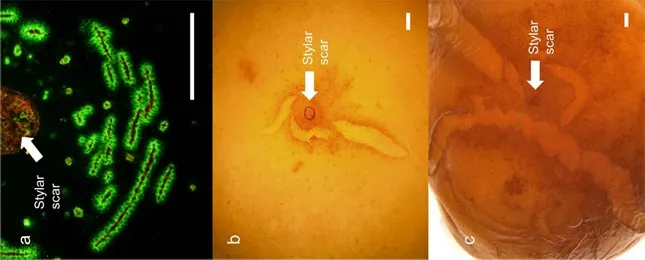
 Immagine 1: La Microfessure nella regione della cicatrice stilare indicizzate mediante infiltrazione di arancio di acridina e microscopia a fluorescenza. b, c Regione cicatriziale stilare dopo periodi di incubazione di 9 h (b) e 24 h (c) in acqua deionizzata. Le microfessurazioni si erano estese fino a diventare crepe macroscopicamente visibili. Barre = 1 mm. Fonte: https://doi.org/10.1038/s41438-019-0161-3.
Sweet cherry fruit cracking is affected by environmental, but also by genetic factors. It is well established that sweet cherry cultivars differ in their susceptibility to cracking. The genetic basis for this difference, however, is unknown. Recently, the first paper that identified QTLs for cracking susceptibility evaluated in segregating populations has been published.
The largest and most robust QTLs were reported for stylar end cracking, which is the most frequent type of cracking. The molecular basis of these QTLs is not clear. In particular, information would be helpful on which of the above events of the Zipper model is the most critical in determining the cracking susceptibility of a particular genotype.
This could help to improve the detected QTLs or even identify the underlying candidate genes. From a practical point of view, this information could eventually help breeders in marker-assisted selection strategies.
The objectives of our study were to phenotype a segregating population of sweet cherries for selected key traits of the Zipper model and for their susceptibility to cracking. We selected the same segregating population of sweet cherry fruit for which the QTLs for cracking susceptibility were established earlier. We focused on cuticle mass per unit area, strain of the cuticle, microcracking and the fruit calcium relations because these represent critical events in the Zipper model.
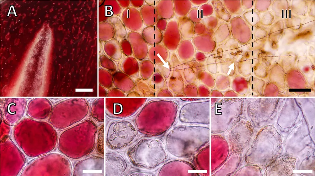
 Immagine 2: Micrografie di crepe macroscopiche (macrocracks) e microscopiche (microcracks) nella buccia della ciliegia "Regina". Fonte: https://doi.org/10.1371/journal.pone.0219794.g004.
Mass of the dewaxed cuticle per unit area and strain release upon cuticle isolation were significantly related to cracking susceptibility in lab or field. Cuticular microcracking in the stylar end region as indexed by infiltration with acridine orange was more severe in susceptible than in tolerant genotypes and significantly correlated with susceptibility to cracking in lab and field. The Ca/dry mass ratio was lower (-8 %) for susceptible than for tolerant genotypes.
Fruit that cracked early had less Ca than those that cracked later. Only the Ca/dry mass ratio of the stylar end region was significantly correlated with cracking susceptibility in the field. Based on stepwise regression analyses microcracking of the cuticle accounted for most of the cracking susceptibilities in field and lab.
The variability in cracking susceptibility accounted for increased when adding mass of the dewaxed cuticle (cracking evaluation in the lab), or when adding the Ca/dry mass ratio in the stylar end region or when entering the strain release on isolation into the model (cracking evaluation in the field).
Our data indicate that microcracking and DCM mass per unit area are the two characteristics that account for most of the crack susceptibility observed in field and lab. Both would be expected based on the Zipper model. Future studies can now focus on phenotyping genotypes for QTLs for these two traits.
Source: Cracking susceptibility of full-sibs of a cross of a cracking tolerant and cracking susceptible sweet cherry: Relation to cuticle characteristics, microcracking and calcium. Knoche M, Grosset-Grange L, Quero-García J, Alletru D, Boutaleb L (2025) Cracking susceptibility of full-sibs of a cross of a cracking tolerant and cracking susceptible sweet cherry: Relation to cuticle characteristics, microcracking and calcium. PLOS ONE 20(1): e0316637. https://doi.org/10.1371/journal.pone.0316637.
Moritz Knoche; José Quero García
Institute for Horticultural Production Systems, Leibniz University Hannover, Germany; INRAE, Biologie du Fruit et Pathologie, Université de Bordeaux, France
Cherry Times - All rights reserved


